
연교 약재 후보 자원, 개나리 (물푸레나무과)의 이화주성 및 성적 이형화현상 연구
This is an open access article distributed under the terms of the Creative Commons Attribution Non-Commercial License (http://creativecommons.org/licenses/by-nc/3.0/) which permits unrestricted non-commercial use, distribution, and reproduction in any medium, provided the original work is properly cited.
Abstract
Forsythia koreana (Rehder) Nakai (family Oleaceae) is a perennial shrub that may be considered as a substitute for F. suspensa and F. viridissima in Korea, which are sources of Forsythiae Fructus. This study was conducted to confirm sexual dimorphism in two F. koreana morphs by analyzing their sexual system (distyly or heterostyly) and pollen viability for effective fruit set.
Floral structure, pollen size, and pollen viability were studied by microscopic analyses. Sepal, petal, and leaf epidermal cells were observed using a scanning electron microscope, which confirmed sexual dimorphism in F. koreana. The thrum flowers were significantly larger and had higher pollen viability than the pin flowers. The micromorphological traits of sepals, petals, and leaves were not significantly different between the two morphs.
F. koreana, with its distyly characteristics, sexual dimorphism, and fruit production, has the potential to be an alternative source of Forsythiae Fructus. However, comprehensive analytical studies of the specific bioactive compounds in this species are essential to confirm their pharmacological suitability compared with those of F. suspensa and F. viridissima, which are widely used.
Keywords:
Forsythia koreana (Rehder) Nakai, Distyly, Floral Dimorphism, Pollen Viability, Scanning Electron Microscope서 언
물푸레나무과 (Oleaceae Hoffmanns. & Link)는 전 세계적으로 약 25 속 600여 종으로 구성되며, 남극지방을 제외한 온대 북부 지역부터 아열대 남부 지역까지 모든 대륙에 분포하는 것으로 알려진 바 있다 (Wallander and Albert, 2000). 이들 중 개나리속 (Forsythia Vahl)은 유라시아의 온대지역, 특히 북·동부 아시아와 동부 유럽에 분포하고 있으며, 약 11 종이 서식하고 있고, 한반도에는 6 종이 서식하는 것으로 보고된 바 있다 (Kim, 1999; Kim, 2018). 개나리속 내 분류군 중 F. europaea Degen & Bald.는 남동부 유럽의 알바니아와 세르비아에 제한적으로 분포하며, 나머지 종들은 극동 지역에 넓게 분포하는 것으로 알려져 있다 (Kim and Kim, 2011).
개나리속은 삭과 (capsule)인 열매, 속이 빈 줄기의 수 (pith), 노란색의 꽃색 등의 형태학적 특징에 의해 물푸레나무과의 다른 속들과 구분된다 (Tae et al., 2005). 또한 개나리속 분류군에서는 이화주성 (distyly, heterostyly)으로 암술대가 수술대보다 긴 형태인 장주화 (pin type, long styles)와 짧은 형태인 단주화 (thrum type, short styles)의 분명한 두 가지 형태를 가지는 생식기관의 이형화현상 (dimorphism)이 관찰된다 (Kim and Kim, 2004; Oh, 2018). 한편 이화주성은 자웅분리수정 (herkogamy)의 한 형태로 자가불화합성 (self-incompatibility)의 특성이 나타나 타가수분을 더욱 촉진하는 식물의 생식 전략으로 알려져 있다 (Barrett, 1990; Lloyd and Webb, 1992).
개나리 [F. koreana (Rehder) Nakai]는 한반도 중부에서 기원하였을 것으로 추정되는 식물로 다년생 관목 (shrub)이고, 높이가 1 m - 3 m에 달하며, 잔가지는 녹색이나 점차 회갈색으로 되고, 피목 (lenticel)이 뚜렷하게 나타나는 특징으로 속 내 다른 분류군과 구별된다 (Lee, 2014; Kim, 2018). 꽃은 노란색이고, 화관 (corolla)은 깊게 4열로 갈라지며, 각 열편은 장타원형이다. 꽃받침은 4개로 갈라지고, 녹색이며, 무모 (glabrous)이다. 잎은 대생하며, 피침형 (lanceolate) 또는 난상장타원형 (ovate-oblong), 예두 (acute)이며, 설저 (cuneate)로 배축면은 무모, 엽연의 중앙 이상에 예거치 (serrate)가 있거나 거의 전연 (entire)이다 (Lee et al., 1982; Lee, 1984; Lee, 2014; Oh, 2018).
개나리속 분류군 중 당개나리 [F. suspensa (Thunb.) Vahl] 및 의성개나리 (F. viridissima Lindl.)는 해열과 해독에 효과를 나타내는 연교 (생약명: Forsythiae Fructus)의 기원종으로 알려진 바 있으며 (Sun et al., 2018), 한국에서는 당개나리의 대체물로 개나리를 사용하는 것으로 보고된 바 있다 (Sun et al., 2018). 한편, 개나리 꽃과 열매에서 추출한 리그난 (lignans)은 항염증 작용에 효과가 있는 것으로 보고된 바 있고 (Lim et al., 2008; Kim et al., 2019), 잎에서 추출한 에센셜 오일 (essential oil)은 천연 식품 방부제로써의 가능성이 제시된 바 있다 (Yang et al., 2015).
전자현미경 (electron microscope)을 활용한 미세형태학적 형질은 현화식물군 내 다양한 분류 계급에서 그 분류학적 유용성이 검증된 바 있다 (Wilkinson, 1979). 개나리속과 미선나무속 (Abeliophyllum Nakai)으로 구성된 개나리족 (Forsythieae H. Taylor ex L. Johnson) 내에서 역시 일부 잎 표피 미세형태학적 형질이 진단형질로서 매우 유용한 것으로 확인되었고 (Song and Hong, 2013), 화분학적 형질은 개나리속 내에서 효과적인 검색표 작성을 위해 활용되었다 (Lee, 1984).
개나리의 성 체계에 관한 연구로는 장주화와 단주화 사이의 화색 및 화형의 차이점을 밝히는 형태 및 분석화학적 연구 (Han and Kim, 1999), 꽃과 잎의 외부형태학적 형질 차이를 밝히는 분류학적 연구가 진행된 바 있다 (Lee et al., 1982). 하지만, 악편, 화판 및 잎 등의 다양한 기관을 미세형태학적으로 자세히 관찰하고, 이화주성에 따라 구별되는 형질을 기재하며, 성 체계에 따른 이형화현상을 비교한 연구는 알려진 바 없다.
본 연구는 개나리의 장주화 및 단주화의 외부형태학적 및 미세형태학적 형질을 관찰하고, 관찰 결과를 바탕으로 두 유형의 꽃에서 나타나는 이형화현상을 기재하여 기초자료를 축적하고자 수행되었다. 나아가 성적 이형화로 인한 형질 차이 및 화분 임성률 비교를 통해 이화주성을 나타내는 성 체계가 자웅이주로의 진화 단계인지 고찰하고자 하였다.
재료 및 방법
1. 연구 재료
개화기 (3월)에 개나리를 수집하여 연구 대상으로 하였으며, 각각 독립된 개체로부터 채집된 장주화와 단주화의 일부를 70% ethanol 용액에 액침표본으로 제작하였고, 나머지 식물체는 석엽표본으로 제작하였다. 사용된 재료 및 표본 정보는 다음과 같다 [pin1; Korea, Chungcheongbuk-do, Cheongju-si, 23. Mar. 2024. Eum 150 (CBU, herbarium acronym), pin2; 26. Mar. 2024. Eum 154 (CBU), thrum1; 26. Mar. 2024. Eum 153 (CBU), thrum2; 26. Mar. 2024. Eum 155 (CBU)].
2. 외부형태
개나리의 생태사진은 스마트폰 (iPhone 12, Apple, Cupertino, CA, USA)으로 촬영하였다. 꽃의 단면을 실체현미경 (S9D, Leika, Wetzlar, Germany)으로 관찰하였고, 생식기관과 열매 및 종자의 외부 형태를 동일한 실체현미경으로 관찰한 후 분석프로그램 (digimizer version 6.3.0, MedCalc Software, Ostend, Belgium)을 이용해 길이를 측정하였다.
3. 화분의 임성 및 크기
화분의 임성률을 측정하기 위해 꽃 한 개당 약 한 개씩, 총 8 개의 약을 1% aniline blue 용액으로 10 분간 염색하고 60 ㎕씩 슬라이드 박편으로 제작하여 동일한 광학현미경으로 관찰하였다. 화분의 극축 길이 및 적도면의 직경을 측정하기 위해 lugol's solution 용액으로 염색하여 광학현미경 (DM750, Leika, Wetzlar, Germany)을 통해 관찰하였다. 크기 측정에 사용한 화분은 임성과 불임성 화분을 모두 포함하여 무작위로 30 개 이상씩을 선별하였다. 또한, 화분의 표벽 두께를 측정하기 위해 acetocarmine 용액으로 염색하여 동일한 광학현미경으로 관찰하였다.
4. 미세구조
영양기관 및 생식기관의 미세형태학적 형질을 관찰하기 위해 임계점 건조기 (SPI-13200J-AB, SPI Supplies, West Chester, PA, USA)를 사용하여 완전 건조시켰으며, 종자는 자연 건조시키는 전처리 과정을 거쳤다. 건조 시료는 직경 9.5 ㎜ 알루미늄 스터브 (01501-BA, SPI Supplies, West Chester, PA, USA) 위에 카본테이프로 고정하여 이온 증착기 (Q150T ES Plus, Quorum Technologies Ltd., Laughton, England)를 이용해 백금 (Pt)으로 45 초간 이온 증착 하였다. 각 기관별로 1 개의 시료를 제작하였으며, 생식기관은 전계방출형 주사전자현미경 (Ultra Plus, Carl Zeiss, Jena, Germany)을 통해 가속전압 3 ㎸, 작동거리 (working distance) 5.0 ㎜ - 5.7 ㎜에서 시료를 관찰하였고, 영양기관과 종자는 주사전자현미경 (EM30+, COXEM, Daejeon, Korea)을 통해 가속전압 5 ㎸, 작동 거리 5.4 - 6.0 ㎜에서 시료를 관찰하였다.
5. 통계분석
연구 결과 데이터는 평균 ± 표준편차 (means ± standard deviation)로 표시하였고, 장주화와 단주화에서 관찰된 정량적 형질 차이는 SPSS 28.0 (SPSS Inc., Chicago, IL, U.S.A.)를 이용하여 Student’s t-test를 진행하였으며, 통계적 유의성을 검증하였다.
결 과
1. 장주화, 단주화의 화부 구조
개나리 (F. koreana)의 꽃은 장주화 (pin) 및 단주화 (thrum)의 두 형태에서 모두 사수성이고, 방사상칭형이며, 합판화이고, 한 개의 암술과 두 개의 수술이 관찰되었다 (Fig. 1A-D).
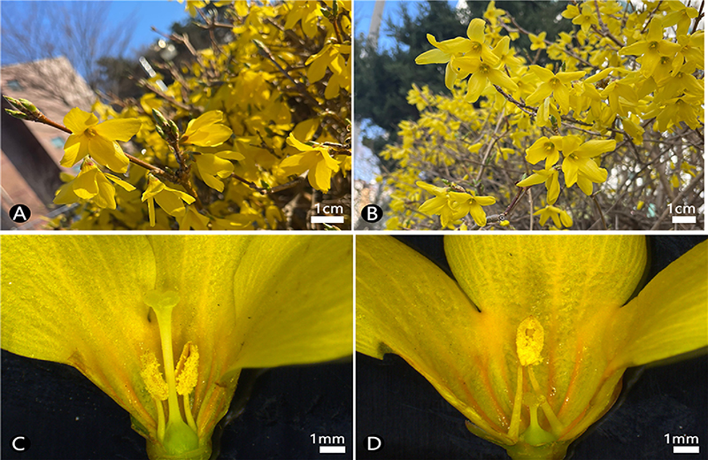
Photographs of the F. koreana. A-B; Habit, C-D; Photographs using stereo microscope (SM), A; Pin flower, B; Thrum flower, C; Longitudinal section of pin flower, D; Longitudinal section of thrum flower.
장주화와 단주화 사이의 화부 구조는 화주와 수술 길이에서 형태적 차이가 명확한 것으로 확인되었다 (Fig. 1C-D). 악편의 길이는 장주화에서 4.50 ± 0.46 ㎜, 단주화에서 4.57 ± 0.37 ㎜로 관찰되었고, 악편의 너비는 장주화에서 2.45 ± 0.26 ㎜, 단주화에서 2.81 ± 0.27 ㎜로 관찰되었다 (Table 1). 화관 열편의 길이는 장주화에서 14.92 ± 0.54 ㎜, 단주화에서 14.40 ± 1.41 ㎜로 관찰되었고, 화관 열편의 너비는 장주화에서 8.26 ± 0.84 ㎜, 단주화에서 8.72 ± 0.40 ㎜로 관찰되었으며, 화관통부의 길이는 장주화에서 4.64 ± 0.18 ㎜, 단주화에서 5.68 ± 0.40 ㎜로 관찰되었다 (Table 1). 화주의 길이는 장주화에서 6.26 ± 1.02 ㎜, 단주화에서 1.83 ± 0.75 ㎜로 관찰되었고, 주두의 길이는 장주화에서 0.57 ± 0.14 ㎜, 단주화에서 0.46 ± 0.09 ㎜로 관찰되었으며, 주두의 너비는 장주화에서 1.01 ± 0.21 ㎜, 단주화에서 1.16 ± 0.15 ㎜로 관찰되었다 (Table 1). 수술의 길이는 장주화에서 4.61 ± 0.29 ㎜, 단주화에서 6.42 ± 0.21 ㎜로 관찰되었다 (Table 1).
2. 화분의 임성 및 크기
화분 임성률은 장주화에서 37.08 ± 15.91 %, 단주화에서 78.59 ± 10.61 %로 관찰되었다 (p < 0.001, Table 2 and Fig. 2A-B). 극축의 길이 (P, polar view)는 장주화에서 28.64 ± 3.98 ㎛, 단주화에서 34.63 ± 2.87 ㎛로 관찰되었고, 적도면의 직경 (E, equatorial view)은 장주화에서 28.72 ± 4.18 ㎛, 단주화에서 36.72 ± 2.29 ㎛로 관찰되었다 (p < 0.001 and Table 2).
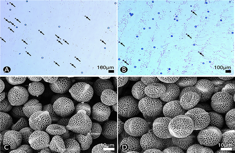
Light microscope (LM) and field emission scanning electron microscope (FE-SEM) micrographs of the pollen grains of F. koreana. A-B; Aniline blue stained pollen viability using LM, C-D; Pollen grains using FE-SEM, A, C; Pin flower, B, D; Thrum flower. Black arrows indicate unviable pollen grains.
장주화와 단주화에서 화분은 모두 단립 (monad)이고, 화분의 형태는 삼공구형 (tricolporate)으로 관찰되었으며, P/E 비율이 장주화에서 1.00 ± 0.07 ㎛, 단주화에서 0.94 ± 0.04 ㎛로 관찰되어 약단구형 (oblate-spheroidal) 내지 구형 (spheroidal)인 것으로 확인되었다. 표벽의 두께는 장주화에서 1.58 ± 0.27 ㎛, 단주화에서 1.53 ± 0.17 ㎛로 관찰되었고, 표면무늬는 망상형 (reticulate)으로 확인되었다 (Table 2 and Fig. 2C-D).
3. 악편, 화판 표피의 미세구조
악편의 외면 표피세포의 모양은 장주화와 단주화에서 모두 등방형 (isodiametric)과 불규칙형 (irregular)이 동시에 관찰되었고, 수층벽 (anticlinal wall)은 직선형 (straight)과 곡선형 (curved)이 동시에 관찰되었으며, 병층벽 (periclinal wall)은 볼록 (convex)하였고, 각피층에는 줄무늬가 관찰되었다 (Fig. 3A-B).
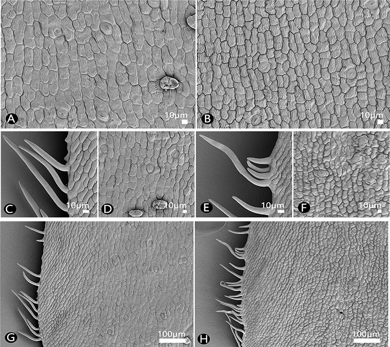
SEM micrographs of the sepal of the F. koreana. A-B; Outer side, C-H; Inner side, A, C-D, G; Pin flower, B, E-F, H; Thrum flower.
악편의 내면 표피세포는 장주화와 단주화에서 모두 등방형과 불규칙형이 동시에 관찰되었고, 수층벽은 직선형과 곡선형이 동시에 관찰되었으며, 병층벽은 볼록하였고, 각피층에는 줄무늬가 관찰되었다 (Fig. 3D, F).
병층벽은 장주화의 외면과 내면에서 모두 약간 볼록한 것으로 관찰되었고, 단주화에서는 볼록한 것으로 관찰되었다 (Fig. 3A, B, D, F). 장주화, 단주화 모두 악편의 가장자리에 단세포 단모 (simple unicellular non-glandular trichome)가 산생하였고, 모용의 표면은 매끈 (smooth)한 것으로 관찰되었다 (Fig. 3C, E, G, H). 장주화, 단주화의 악편에서 기공복합체는 외면과 내면 모두에서 기공이 관찰되었고, 기공복합체의 형태는 부재형 (anomocytic type)으로 관찰되었다 (Fig. 3A, B, D, F).
화주의 길이는 장주화가 단주화에 비하여 긴 것으로 관찰되었으며 (Fig. 4A, D), 주두의 너비는 단주화가 장주화에 비하여 크게 나타난 것으로 관찰되었고 (Fig. 4B, E), 장주화, 단주화 화주의 표피에서 모두 기공의 형태가 관찰되었다 (Fig. 4C, F).
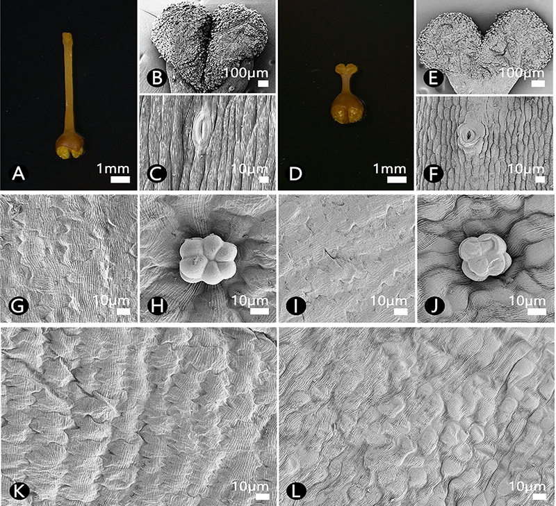
SM and SEM micrographs of floral details of the F. koreana. A-F; Pistil, A, D; Pistil (SM image), B, E; Stigma, C, F; Style (SEM image), G-J; Outer side of the petal, K-L; Inner side of the petal, H, J; Glandular trichome, A-C, G - H, K; Pin flower, D-F, I-J, L; Thrum flower.
화판의 외면 표피세포는 장주화와 단주화에서 모두 등방형의 직사각형 (rectangle)으로 관찰되었고, 수층벽은 파상형 (undulate)으로 관찰되었으며, 병층벽은 평평 (flat)했고, 각피층에는 줄무늬가 관찰되었다 (Fig. 4G, I). 화판의 내면 표피세포는 장주화에서 등방형의 직사각형으로 관찰되었고, 단주화에서 불규칙형으로 관찰되었다 (Fig. 4K-L). 장주화에서 화판의 내면 수층벽은 파상형이었으며, 병층벽은 약간 볼록했고, 각피층에는 줄무늬가 관찰되었다 (Fig. 4K). 단주화에서 화판의 내면 수층벽은 직선형 또는 곡선형이었으며, 병층벽은 장주화와 동일하게 약간 볼록했고, 각피층에는 줄무늬가 관찰되었다 (Fig. 4L). 장주화, 단주화 모두 화판의 외면과 내면에서 방패형 선모 (subsessile/peltate glandular trichome)가 관찰되었으며, 두상 (capitate)의 구성세포는 3개에서 6개로 나타났다 (Fig. 4H, and J).
4. 잎 표피의 미세구조
장주화와 단주화의 잎은 기공복합체가 배축면에만 존재하는 이면기공형 (hypostomatic type)으로 관찰되었다 (Fig. 5A-B, G-H). 장주화와 단주화의 기공복합체 유형은 부재형으로 관찰되었으며 (Fig. 5G-H), 공변세포의 크기는 장주화에서 21.63 ± 2.85 × 14.60 ± 2.39 (L × W) ㎛, 단주화에서 21.82 ± 2.12 × 15.36 ± 1.45 ㎛로 관찰되었다 (Table 4).
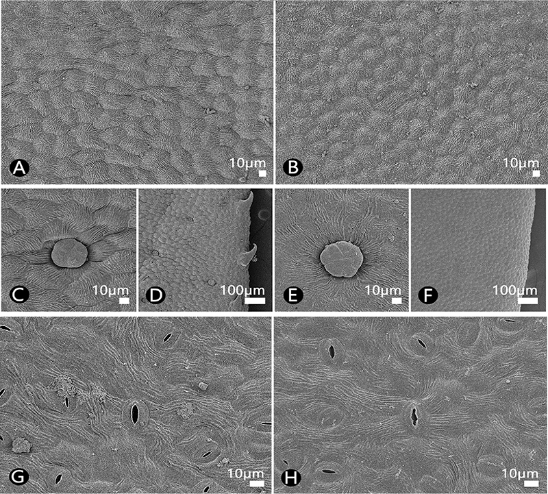
SEM micrographs of the leaf adaxial and the abaxial surface of the F. koreana. A-F; Adaxial surface, G-H; Abaxial surface, A, C-D, G; Pin flower, B, E-F, H; Thrum flower.
잎의 향축면 표피세포는 장주화와 단주화에서 모두 등방형으로 관찰되었고, 수층벽은 직선형과 곡선형이 동시에 관찰되었으며, 병층벽은 볼록하였고, 각피층에는 줄무늬가 관찰되었다 (Fig. 5A-B). 장주화, 단주화 모두 잎의 향축면에서 방패형 선모가 관찰되었으며, 두상의 구성세포는 4개에서 8개로 확인되었다 (Fig. 5C, E). 장주화에만 잎의 향축면에서 가장자리에 짧은 단세포 단모가 나타났고, 모용의 표면은 매끈한 것으로 관찰되었다 (Fig. 5D, F).
잎의 배축면 표피세포는 장주화와 단주화에서 모두 불규칙형으로 관찰되었고, 수층벽은 심파상형 (sinuate)이 관찰되었으며, 병층벽은 약간 볼록하였고, 각피층에는 줄무늬가 관찰되었다 (Fig. 5G-H). 장주화, 단주화 모두 잎의 배축면에서 방패형 선모가 관찰되었으며, 세포는 4개에서 6개로 확인되었다.
5. 종자 표피세포의 미세구조
열매는 삭과로 (Fig. 6A), 종피 색은 갈색으로 관찰되었고, 모양은 신장형 (reniform)에서 타원체형 (ellipsoid)으로 확인되었다 (Fig. 6B-C). 종자의 크기는 길이 1.56 ± 0.19 ㎜, 너비 0.66 ± 0.09 ㎜로 측정되었다 (Table 5). 종자 표면무늬는 불규칙한 다각형으로 이루어진 망상형 (reticulate)으로 관찰되었고, 망벽 (muri)의 두께는 9.72 ± 1.32 ㎛, 망강 (lumen)의 면적은 812.49 ± 243.02 ㎛2로 측정되었다 (Table 5 and Fig. 6D-E).
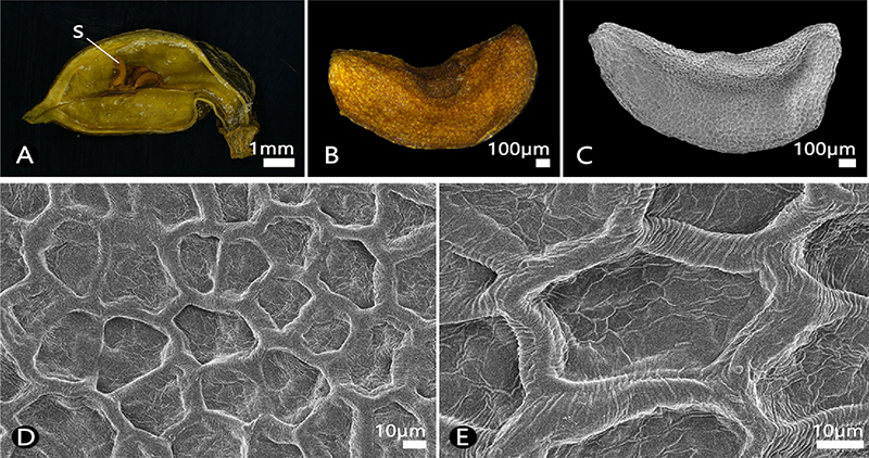
SM and SEM micrographs of the seed of the F. koreana. A; Fruit (SM image), B; Seed (SM image), C; Seed (SEM image), D-E; Detailed seed surface (SEM image). S; Seed.
고 찰
본 연구에서 개나리의 화주 길이는 장주화에서 단주화보다 길게 나타났고, 수술의 길이는 단주화에서 장주화보다 길게 나타났으며, 이는 통계적으로 매우 유의미한 형태적 차이로 확인되었다 (p < 0.001).
이전 연구에서 화주의 길이는 장주화에서 6.45 ± 0.09 ㎜, 단주화에서 1.89 ± 0.07 ㎜, 수술대의 길이는 장주화에서 2.34 ± 0.06 ㎜, 단주화에서 4.40 ± 0.11 ㎜로 관찰되어 (Lee et al., 1982), 본 연구 결과와 일치하는 것으로 확인되었다. 다만, 악편의 길이는 장주화에 비해 단주화에서 약간 크게 나타나, 기존의 연구 (Lee et al., 1982)와는 상이한 것으로 확인되었다. 그러나, 이는 유의확률 50%로 추정된 값으로 본 연구에서 악편의 길이는 장주화와 단주화 사이의 차이가 통계적으로 유의미하지 않다고 판단된다 (p < 0.5).
추후 다양한 지역에 분포하는 대상 분류군으로 시료의 확장을 통해서, 악편의 길이 차이가 환경적 요인에 의한 변이일지, 이화주성에 따른 이형화현상일지 분류학적, 생태학적 연구가 필요할 것으로 생각된다.
화분의 임성은 단주화가 장주화보다 높게 확인되었고, 화분의 크기 또한 단주화가 장주화보다 크게 관찰되어 통계적으로 매우 유의미한 차이를 나타내는 것으로 확인되었다 (p < 0.001).
다양한 현화식물에서 화분관이 신장할 때는 충분한 양의 에너지가 필요하며, 화주의 길이와 화분의 크기 사이에는 양의 상관관계가 있다고 제안된 바 있다 (Lee, 1978; Ganders, 1979; Cruden and Lyon, 1985). 또한, 이화주성의 성 체계를 지니는 식물 대부분에서 단주화의 화분이 장주화에 비해 크기가 크게 관찰되는 것으로 알려진 바 있다 (Pailler and Thompson, 1997).
본 연구에서 관찰된 화분의 크기 차이는 단주화의 화분이 장주화의 긴 화주를 따라 신장하는 과정에서 장주화의 화분보다 더 많은 에너지를 필요로 하기 때문에 단주화의 화분 크기가 더 크게 관찰되는 것으로 판단하였다.
한편, 양성화로부터 단성화로 이어지는 성의 분리가 현화식물군 내에서의 진화적 경향성이며, 이때 성적 이형화현상을 동반하는 것으로 제안된 바 있다 (Ashman, 2009).
본 연구의 결과, 화분의 크기가 크고, 임성률이 높은 단주화가 장주화에 비해 웅성의 기능에 충실한 것으로 해석하였고, 본 분류군에서 관찰되는 이화주성 또한 자웅이주로의 진화 단계 중 중간 형태일 것으로 추론하였다. 추후 이화주성에 따른 열매의 결실률 비교를 통해 단주화 및 장주화의 웅성 및 자성의 기능과 성의 분화에 대한 연구가 수행되어야 할 것으로 생각된다.
악편의 표피세포의 형태는 장주화와 단주화에서 큰 차이를 확인할 수 없었다. 다만, 병층벽은 외면과 내면에서 모두 단주화가 장주화보다 볼록한 것으로 관찰되었다. 장주화, 단주화의 악편에서 기공복합체는 양면 모두에 존재했으며, 기공복합체의 형태는 부재형으로 관찰되었다. 또한 장주화, 단주화 모두 악편의 가장자리에 단세포 단모가 산생하였고, 모용의 표면은 매끈한 것으로 관찰되어, 무모인 악편으로 기재한 이전의 결과 (Lee, 2014)와 상이하였다. 추후 다양한 서식지의 분류군으로 시료의 확장을 통해 악편의 모용 유무가 서식지에 따른 변이의 양상인지 보다 면밀한 분류학적 연구가 필요할 것으로 생각된다.
화주의 길이는 항상 장주화가 단주화에 비하여 긴 것으로 관찰되었고, 주두의 너비는 단주화가 장주화에 비하여 큰 것으로 관찰되었으며, 장주화 및 단주화 화주의 표피에서 모두 기공의 형태가 관찰되었다. 선행된 연구에서 물푸레나무과에 속하며 이화주성이 나타나는 미선나무 (Abeliophyllum distichum Nakai)는 항상 단주화보다 장주화에서 화주의 길이가 긴 것으로 확인되었고, 기공형 밀선 (stomata-like nectary)이 화주 표피에서 관찰된 바 있다 (Hong and Han, 2002).
본 연구에서 관찰된 기공의 형태가 기공형 밀선일 것으로 판단되며, 추후 화부구조에서 관찰되는 기공과 유사한 구조가 어떤 역할을 수행하는지 생리학적, 생태학적 연구가 진행되어야 할 것으로 생각된다.
장주화, 단주화 모두 화판의 양면에서 방패형 선모가 관찰되었으며, 두상의 구성세포는 3개에서 6개로 관찰되었고, 화판의 외면 표피세포는 장주화와 단주화에서 차이가 관찰되지 않았다. 화판의 내면 표피세포는 장주화에서 등방형의 직사각형으로 관찰되었고, 수층벽은 파상형으로 관찰되었다. 반면, 단주화에서 화판 내면의 표피세포가 불규칙형으로 관찰되었으며, 수층벽은 직선형 또는 곡선형으로 관찰되어 장주화와 차이를 보였다. 개나리속의 일부 분류군은 곤충이 수분을 매개하는 것으로 알려진 바 있으나 (Chung et al., 2013), 평평한 세포 표면을 가진 꽃은 원추형 세포 (conical cell)를 가진 꽃보다 곤충의 방문이 적은 것으로 알려진 바 있다 (Glover and Martin, 1998). 한편, 노란색 꽃은 보통 벌 뿐만 아니라 새도 수분을 매개하고, 꽃잎에 특정한 UV 패턴을 가지고 있으며, 이는 벌과 새의 이목을 끄는 것으로 알려진 바 있다 (Papiorek et al., 2016).
따라서 개나리의 화판 표피세포의 형태보다는, 화판에서 나타나는 노란색 색소가 수분매개자를 유도하는 현상과 연관되었을 것으로 판단하였다. 다만, 개나리에서 수분매개자의 종류는 보고된 것이 전무하기 때문에, 추후 수분매개자와 연관하여 생태학적 연구가 수행되어야 할 것으로 생각된다.
장주화와 단주화의 잎은 기공복합체가 배축면에만 존재하는 이면기공형으로 관찰되었고, 기공복합체 유형은 부재형으로 관찰되었으며, 공변세포의 크기는 장주화에서 21.63 ± 2.85 × 14.60 ± 2.39 ㎛, 단주화에서 21.82 ± 2.12 × 15.36 ± 1.45 ㎛로 관찰되었다. 잎의 향축면 표피세포는 장주화와 단주화에서 모두 등방형으로 관찰되었고, 수층벽은 직선형과 곡선형이 동시에 관찰되었으며, 병층벽은 볼록하였고, 각피층에는 줄무늬가 관찰되었다.
장주화, 단주화 모두 잎의 향축면에서 방패형 선모가 관찰되었으며, 두상의 구성세포는 4개에서 8 개로 확인되었다. 장주화에만 잎의 향축면에서 가장자리에 짧은 단세포 단모가 나타났고, 모용의 표면은 매끈한 것으로 관찰되었다. 모용의 경우 발달의 이른 시기에 퇴화되거나 성숙 직후 탈락되는 경우가 보고된 바 있기에 (Metcalf and Chalk, 1979), 추후 다양한 지역 및 채집시기를 달리하여 시료를 확장하고, 잎의 모용 유무가 변이의 양상인지, 이화주성에 따른 성적 이형화현상인지, 보다 면밀한 분류학적 연구가 필요할 것으로 생각된다.
본 연구에서 종자의 크기는 1.56 ± 0.19 × 0.66 ± 0.09 ㎜로 측정되었다. 이전 연구에서는 4.67 - 5.33 × 1.44 - 1.83 ㎜로 기재된 바 있어 (KNA, 2017), 본 연구 결과와 매우 상이한 것으로 확인되었다. 완전 성숙 후 열개한 삭과 내 종자의 크기를 측정하였으나 이들 크기의 차이가 지역적 혹은 성장적 변이 일지 추후 다양한 개체를 대상으로 연구를 진행해야 할 것이다. 또한 성숙 후에도 불완전 결실일 경우를 배제할 수 없어 추후 종자 결실률 및 활력 검정 비교 연구를 통해 보다 명확한 결실의 차이를 제시해야 할 것이다.
종자의 표면무늬는 불규칙한 다각형으로 구성된 망상형으로 관찰되었다. 선행된 연구에서 개나리의 종자 표면무늬는 구릉상 (colliculate) 또는 망상 (reticulate)으로 기재된 바 있다 (KNA, 2017; Ghimire et al., 2018). 본 연구를 통해 개나리의 종피는 구릉상이 아닌 망상으로 명확히 기재하는 바이다.
종자의 크기는 위도, 고도, 온도 등의 환경적 변수에 적응한 결과 나타나는 변이뿐만 아니라, 성장 시기에 따른 성장적 변이도 영향을 미칠 수 있다고 보고되어 있다 (Ramírez-Valiente, 2009). 반면, 종자의 표면무늬는 환경적 영향을 비교적 적게 받아 분류군을 구별하는 매우 유용한 형질로 알려져 있다 (Barthlott, 1984). 이에, 추후 전략적으로 다양한 서식지 및 성숙 시기별 종자 수집을 통해 장주화, 단주화의 종자 형태 크기 및 종피 패턴의 변이 차이를 규명할 필요가 있겠다.
의성지역 개나리 수집 자원의 결실 유형 연구에서 의성개나리는 장주화만 관찰되었으며, 상당량의 열매를 맺는 자가화합성이라고 제시된 바 있다 (Lee, 1984; Kim and Park, 2022). 또한 해당 연구에서 Leaf I 유형을 나타낸 개나리 계통은 모두 무결실로 관찰된 바 있다 (Kim and Park, 2022). 하지만 본 연구를 통해 개나리는 자가불화합성을 나타내지만, 장주화와 단주화 개체 사이의 유성생식을 통해 열매 생산이 가능함을 확인하였다. 또한 개나리는 자가불화합성을 통해 자가수정에서 발생하는 근교약세 현상과 낮은 유전적 다양성의 발생을 방지할 수 있어, 높은 유전적 다양성을 유지할 수 있을 것이라 판단하였다.
한편, 계통분류학적 및 진화적으로 유연관계가 가까운 근연 분류군의 경우, 유사한 효능을 나타낼 것이라는 예견적 가치 (predictive value)에 따라 계통수 기반 대체 의약 자원 탐색 연구가 진행되고 있다 (Ernst et al., 2016; Seo et al., 2024). 개나리 역시 의성개나리와 매우 가까운 근연관계로 단계통군 (monophyly)을 나타내는 바 (Ha et al., 2018), 유성 생식을 통한 효과적인 개나리 열매 생산 및 재배 시스템 구축을 통해 연교의 대체작물로 활용이 가능할지 고민해볼 필요가 있겠다.
결론적으로, 본 연구를 통해 개나리의 이화주성 및 외부형태학적 및 미세형태학적 형질의 성적 이형화현상을 보다 명확히 확인하였으며, 장주화와 단주화의 유성생식을 통해 삭과인 열매와 종자를 생성하는 것을 확인하였다. 이를 통해 연교 약재의 기원종인 당개나리, 의성개나리의 대체품으로써의 가능성을 제안할 수 있었으며, 추후 열매 재배 특성, 약효 및 활용에 중점을 둔 연구가 수행되어야 할 것으로 생각된다.
Acknowledgments
본 연구는 정부(과학기술정보통신부)의 재원으로(Grant No. RS-2023-00208589) 한국연구재단의 지원에 의해 이루어진 결과로 이에 감사드립니다.
References
-
Ashman TL. (2009). Sniffing out patterns of sexual dimorphism in floral scent. Functional Ecology. 23:852-862.
[https://doi.org/10.1111/j.1365-2435.2009.01590.x]

-
Barrett SCH. (1990). The evolution and adaptive significance of heterostyly. Trends in Ecology and Evolution. 5:144-148.
[https://doi.org/10.1016/0169-5347(90)90220-8]

- Barthlott W. (1984). Microstructural features of seed surfaces. In Heywood VH. and Moore DM. (eds.). Current concepts in plant taxonomy. Academic Press. London, England. p.95-105.
-
Chung MY, Chung JM, López-Pujol J, Park SJ and Chung MG. (2013). Genetic diversity in three species of Forsythia (Oleaceae) endemic to Korea: Implications for population history, taxonomy, and conservation. Biochemical Systematics and Ecology. 47:80-92.
[https://doi.org/10.1016/j.bse.2012.11.005]

-
Cruden RW and Lyon DL. (1985). Correlations among stigma depth, style length, and pollen grain size: Do they reflect function or phylogeny?. Botanical Gazette. 146:143-149.
[https://doi.org/10.1086/337509]

-
Ernst M, Saslis-Lagoudakis CH, Grace OM, Nilsson N, Simonsen HT, Horn JW and Rønsted N. (2016). Evolutionary prediction of medicinal properties in the genus Euphorbia L. Scientific Reports. 6:30531. (cited by 2024 Dec. 20)
[https://doi.org/10.1038/srep30531]

-
Ganders FR. (1979). The biology of heterostyly. New Zealand Journal of Botany. 17:607-635.
[https://doi.org/10.1080/0028825X.1979.10432574]

-
Ghimire B, Jeong MJ, Suh GU, Heo K and Lee CH. (2018). Seed morphology and seed coat anatomy of Fraxinus, Ligustrum and Syringa(Oleeae: Oleaceae) and its systematic implications. Nordic Journal of Botany. 36:e01866. (cited by 2024 Dec. 20)
[https://doi.org/10.1111/njb.01866]

-
Glover BJ and Martin C. (1998). The role of petal cell shape and pigmentation in pollination success in Antirrhinum majus. Heredity. 80:778-784.
[https://doi.org/10.1038/sj.hdy.6883450]

-
Ha YH, Kim C, Choi K and Kim JH. (2018). Molecular phylogeny and dating of Forsythieae(Oleaceae) provide insight into the Miocene history of Eurasian temperate shrubs. Frontiers in Plant Science. 9:99. (cited by 2024 Dec. 20)
[https://doi.org/10.3389/fpls.2018.00099]

- Han IS and Kim JG. (1999). Characteristics of flowers and flowering between distyly in Korean golden-bells(Forsythia koreana Nak.). Horticulture Environment and Biotechnology. 40:769-771.
-
Hong SP and Han MJ. (2002). The floral dimorphism in the rare endemic plant, Abeliophyllum distichum Nakai(Oleaceae). Flora-Morphology, Distribution, Functional Ecology of Plants. 197:317-325.
[https://doi.org/10.1078/0367-2530-00047]

-
Kim DK and Kim JH. (2004). Numerical taxonomy of tribe Forsythieae(Oleaceae) in Korea. Korean Journal of Plant Taxonomy. 34:189-203.
[https://doi.org/10.11110/kjpt.2004.34.3.189]

-
Kim DK and Kim JH. (2011). Molecular phylogeny of tribe Forsythieae(Oleaceae) based on nuclear ribosomal DNA internal transcribed spacers and plastid DNA trnL-F and matK gene sequences. Journal of Plant Research. 124:339-347.
[https://doi.org/10.1007/s10265-010-0383-9]

-
Kim KJ. (1999). Molecular phylogeny of Forsythia(Oleaceae) based on chloroplast DNA variation. Plant Systematics and Evolution. 218:113-123.
[https://doi.org/10.1007/BF01087039]

- Kim KJ. (2018). Oleaceae Hoffmanns. & Link. In Flora of Korea Editorial Committee (eds.). The genera of vascular plants of Korea. Hongreung Publishing Co. Seoul, Korea. p.1124-1129.
-
Kim KS and Park SH. (2022). Differences of morphological characteristics and lignan components of leaves by distyly and fruiting habits in Uiseong local germplasm of Forsythia spp. Korean Journal of Medicinal Crop Science. 30:89-98.
[https://doi.org/10.7783/KJMCS.2022.30.2.89]

-
Kim TW, Shin JS, Chung KS, Lee YG, Baek NI and Lee KT. (2019). Anti-inflammatory mechanisms of Koreanaside A, a lignan isolated from the flower of Forsythia koreana, against LPS-induced macrophage activation and DSS-induced colitis mice: the crucial role of AP-1, NF-κB, and JAK/STAT signaling. Cells. 8:1163. (cited by 2024 Dec. 20)
[https://doi.org/10.3390/cells8101163]

- Korea National Arboretum(KNA). (2017). Seed atlas of Korea. Sumeunkil Publishing Co. Seoul, Korea. p.640.
-
Lee ST, Kim MY and Hong SP. (1982). A taxonomic study of Forsythia koreana Nakai and F. saxatilis Nakai. Korean Journal of Plant Taxonomy. 12:51-62.
[https://doi.org/10.11110/kjpt.1982.12.2.051]

-
Lee ST. (1978). A factor analysis study of the functional significance of angiosperm pollen. Systematic Botany. 3:1-19.
[https://doi.org/10.2307/2418528]

-
Lee ST. (1984). A systematic study of Korean Forsythia species. Korean Journal of Plant Taxonomy. 14:87-107.
[https://doi.org/10.11110/kjpt.1984.14.2.087]

- Lee TB. (2014). Coloured Flora of Korea. Hyangmunsa. Seoul, Korea. p.426-428.
-
Lim H, Lee JG, Lee SH, Kim YS and Kim HP. (2008). Anti-inflammatory activity of phylligenin, a lignan from the fruits of Forsythia koreana, and its cellular mechanism of action. Journal of Ethnopharmacology. 118:113-117.
[https://doi.org/10.1016/j.jep.2008.03.016]

-
Lloyd DG and Webb CJ. (1992). The evolution of heterostyly. In Barrett SCH. (eds.). Evolution and function of heterostyly. Monographs on theoretical and applied genetics. Springer. Berlin, Germany. p.151-178.
[https://doi.org/10.1007/978-3-642-86656-2_6]

- Metcalfe CR and Chalk L. (1979). Anatomy of the dicotyledons. Systematic anatomy of leaf and stem, with a brief history of the subject. Clarendon Press. Oxford, England. p.40-53.
- Oh SH. (2018). Forsythia Vahl. In Flora of Korea Editorial Committee (eds.). Flora of Korea, Asteridae: Loganiaceae to Oleaceae. Junghaengsa. Seoul, Korea. p.136-138.
-
Pailler T and Thompson JD. (1997). Distyly and variation in heteromorphic incompatibility in Gaertnera vaginata(Rubiaceae) endemic to La Reunion Island. American Journal of Botany. 84:315-327.
[https://doi.org/10.2307/2446005]

-
Papiorek S, Junker RR, Alves‐dos‐Santos I, Melo GAR, Amaral‐Neto LP, Sazima M, Wolowski M, Freitas L and Lunau K. (2016). Bees, birds and yellow flowers: Pollinator‐dependent convergent evolution of UV patterns. Plant Biology. 18:46-55.
[https://doi.org/10.1111/plb.12322]

-
Ramírez-Valiente JA, Valladares F, Gil L and Aranda I. (2009). Population differences in juvenile survival under increasing drought are mediated by seed size in cork oak(Quercus suber L.). Forest Ecology and Management. 257:1676-1683.
[https://doi.org/10.1016/j.foreco.2009.01.024]

-
Seo YS, Song JH, Kim HS, Nam HH, Yang S, Choi G, Chae SW, Lee J, Jung B, Kim JS and Park I. (2024). An integrative study of Scrophularia takesimensis Nakai in an ovalbumin-induced murine model of asthma: the effect on T helper 2 cell activation. Pharmaceutics. 16:529. (cited by 2024 Dec. 20)
[https://doi.org/10.3390/pharmaceutics16040529]

-
Song JH and Hong SP. (2013). The systematic consideration of leaf epidermal microstructure in the tribe Forsythieae and its related genera(Oleaceae). Korean Journal of Plant Taxonomy. 43:118-127.
[https://doi.org/10.11110/kjpt.2013.43.2.118]

-
Sun L, Rai A, Rai M, Nakamura M, Kawano N, Yoshimatsu K, Suzuki H, Kawahara N, Saito K and Yamazaki M. (2018). Comparative transcriptome analyses of three medicinal Forsythia species and prediction of candidate genes involved in secondary metabolisms. Journal of Natural Medicines. 72:867-881.
[https://doi.org/10.1007/s11418-018-1218-6]

- Tae KH, Kim DK and Kim JH. (2005). Genetic variations and phylogenetic relationships of tribe Forsythieae(Oleaceae) based on RAPD analysis. Korean Journal of Plant Resources. 8:135-144.
-
Wallander E and Albert VA. (2000). Phylogeny and classification of Oleaceae based on rps16 and trnL-F sequence data. American Journal of Botany. 87:1827-1841.
[https://doi.org/10.2307/2656836]

- Wilkinson HP. (1979). The plant surface(mainly leaf). In Metcalfe CR. and Chalk L. (eds.). Anatomy of the dcotyledons. Clarendon Press. Oxford, England. p.97-165.
-
Yang XN, Khan I, and Kang SC. (2015). Chemical composition, mechanism of antibacterial action and antioxidant activity of leaf essential oil of Forsythia koreana deciduous shrub. Asian Pacific Journal of Tropical Medicine. 8:694-700.
[https://doi.org/10.1016/j.apjtm.2015.07.031]

